AI & ML Models
0.0 (0 reviews) • 0 downloads
Foetal Segmentation using Image in Python Projects
र 1000
Foetal Segmentation using Image in Python Projects
Technical Details
Domain : Python
Database : Sqlite
Tools : Anaconda
Run Tools: VS Code
Database : Sqlite
Tools : Anaconda
Run Tools: VS Code
Secure Payment
Instant Download
GST Invoice
24/7 Support
About This Product
Foetal Segmentation using Image in Python Projects
Abstract
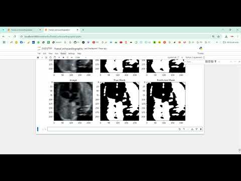
Foetal segmentation in medical imaging is a crucial task for prenatal diagnosis, growth monitoring, and early detection of abnormalities. The project Foetal Segmentation using Image in Python Projects aims to develop an automated system that segments the foetus from ultrasound or MRI images using image processing and deep learning techniques. Python is chosen as the development platform due to its extensive libraries for medical image analysis, machine learning, and visualization, including OpenCV, NumPy, TensorFlow, Keras, and SimpleITK. The system preprocesses input images to remove noise, enhance contrast, and normalize dimensions, then applies convolutional neural networks (CNNs) or U-Net architectures for pixel-level segmentation. Accurate segmentation helps obstetricians and radiologists analyze foetal structures, monitor development, and plan medical interventions.
Existing System
Existing methods for foetal segmentation largely depend on manual annotation and analysis by radiologists or sonographers. Manual segmentation is time-consuming, labor-intensive, and prone to inter-observer variability, which can affect diagnostic accuracy. Some semi-automated approaches use edge detection, thresholding, or region-growing algorithms to segment the foetus, but these techniques often fail in low-contrast or noisy ultrasound images. Traditional image processing methods are also sensitive to foetal movement, maternal tissue interference, and varying imaging conditions, which limits their reliability and scalability. Consequently, there is a strong need for automated, accurate, and robust segmentation systems.
Proposed System
The proposed system introduces a Python-based framework for automatic foetal segmentation using deep learning models. Preprocessing steps include noise reduction, contrast enhancement, image normalization, and resizing to standard dimensions. The system uses U-Net, SegNet, or other CNN-based architectures trained on labeled foetal image datasets to perform precise pixel-level segmentation. Data augmentation techniques such as rotation, flipping, and scaling are applied to improve model generalization and robustness across diverse imaging conditions. The system outputs segmented foetal regions, which can be overlaid on the original images for visualization. Performance is evaluated using metrics such as Dice coefficient, Intersection over Union (IoU), precision, and recall. This approach provides accurate, consistent, and scalable foetal segmentation, supporting prenatal care, growth assessment, and early detection of developmental abnormalities.
Abstract
Foetal segmentation in medical imaging is a crucial task for prenatal diagnosis, growth monitoring, and early detection of abnormalities. The project Foetal Segmentation using Image in Python Projects aims to develop an automated system that segments the foetus from ultrasound or MRI images using image processing and deep learning techniques. Python is chosen as the development platform due to its extensive libraries for medical image analysis, machine learning, and visualization, including OpenCV, NumPy, TensorFlow, Keras, and SimpleITK. The system preprocesses input images to remove noise, enhance contrast, and normalize dimensions, then applies convolutional neural networks (CNNs) or U-Net architectures for pixel-level segmentation. Accurate segmentation helps obstetricians and radiologists analyze foetal structures, monitor development, and plan medical interventions.
Existing System
Existing methods for foetal segmentation largely depend on manual annotation and analysis by radiologists or sonographers. Manual segmentation is time-consuming, labor-intensive, and prone to inter-observer variability, which can affect diagnostic accuracy. Some semi-automated approaches use edge detection, thresholding, or region-growing algorithms to segment the foetus, but these techniques often fail in low-contrast or noisy ultrasound images. Traditional image processing methods are also sensitive to foetal movement, maternal tissue interference, and varying imaging conditions, which limits their reliability and scalability. Consequently, there is a strong need for automated, accurate, and robust segmentation systems.
Proposed System
The proposed system introduces a Python-based framework for automatic foetal segmentation using deep learning models. Preprocessing steps include noise reduction, contrast enhancement, image normalization, and resizing to standard dimensions. The system uses U-Net, SegNet, or other CNN-based architectures trained on labeled foetal image datasets to perform precise pixel-level segmentation. Data augmentation techniques such as rotation, flipping, and scaling are applied to improve model generalization and robustness across diverse imaging conditions. The system outputs segmented foetal regions, which can be overlaid on the original images for visualization. Performance is evaluated using metrics such as Dice coefficient, Intersection over Union (IoU), precision, and recall. This approach provides accurate, consistent, and scalable foetal segmentation, supporting prenatal care, growth assessment, and early detection of developmental abnormalities.
Customer Reviews (0)
No reviews yet. Be the first!
Related Products
⭐ Featured

AI & ML Models
Zomato Restaurant Reviews Sentimental Analyzer in Python Projects
Zomato Restaurant Reviews Sentimental Analyzer in Python Projects
र 1000
⭐ Featured
⭐ Featured
AI & ML Models
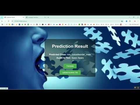
Voice Disorder Prediction using Audio Dataset in Python Projects
Voice Disorder Prediction using Audio Dataset in Python Projects
र 1000
AI & ML Models
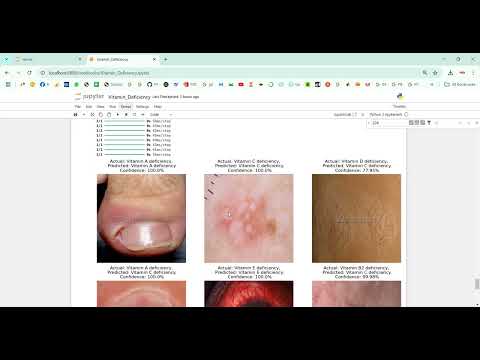
Vitamin Deficiency Detection Using Image Processing in Python Projects
Vitamin Deficiency Detection Using Image Processing in Python Projects
र 1000