AI & ML Models
0.0 (0 reviews) • 0 downloads
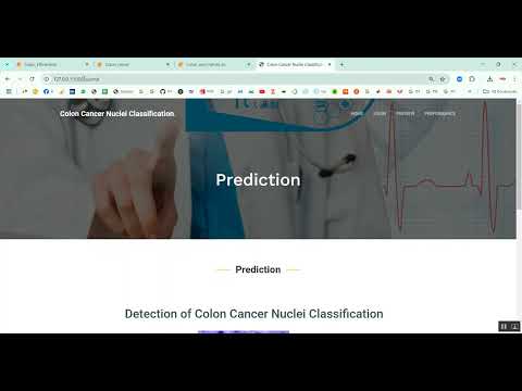
Colon Cancer Nuclei Classification in Python Projects
र 1000
Colon Cancer Nuclei Classification in Python Projects
Technical Details
Domain : Python
Database : Sqlite
Tools : Anaconda
Run Tools: VS Code
Database : Sqlite
Tools : Anaconda
Run Tools: VS Code
Secure Payment
Instant Download
GST Invoice
24/7 Support
About This Product
Colon Cancer Nuclei Classification in Python Projects
Abstract
Colon cancer is one of the most common and life-threatening cancers worldwide. Accurate and early diagnosis plays a crucial role in effective treatment and patient survival. The project titled Colon Cancer Nuclei Classification in Python Projects focuses on building an automated classification system that identifies cancerous cell nuclei from histopathological images of colon tissue. Python is used as the development platform due to its powerful libraries for image processing and deep learning, such as OpenCV, NumPy, TensorFlow, Keras, and Scikit-learn. The system processes microscopic images, segments nuclei regions, extracts relevant features, and classifies them as benign or malignant using Convolutional Neural Networks (CNN). This automated diagnostic tool enhances pathological analysis by increasing detection accuracy, reducing manual effort, and supporting medical experts in clinical decision-making.
Existing System
The existing system for colon cancer diagnosis mainly relies on manual examination of histopathology slides by pathologists. This process is time-consuming, labor-intensive, and subject to human error due to fatigue or variations in expertise. Traditional computer-aided diagnosis (CAD) systems use hand-crafted features like shape, size, and texture of cell nuclei combined with machine learning classifiers. However, these systems often lack accuracy because they depend on manual feature extraction, which may not capture complex patterns present in cancerous cells. Additionally, variations in staining quality, image noise, and overlapping nuclei further reduce the performance of traditional approaches. As a result, current methods are limited in scalability and consistency, highlighting the need for improved automated solutions.
Proposed System
The proposed system introduces a Python-based deep learning approach for colon cancer nuclei classification using histopathological images. The input images are first preprocessed through normalization, denoising, and stain enhancement to improve visual clarity. Nuclei segmentation is performed using thresholding or watershed algorithms to isolate regions of interest. A Convolutional Neural Network (CNN) is then applied to automatically learn complex spatial and morphological features from the segmented images. Transfer learning models such as ResNet50, VGG16, or InceptionV3 can also be employed to improve classification performance and reduce training time. The system is trained using publicly available datasets such as the MoNuSeg or NCT-CRC-HE-100K dataset. Performance is evaluated using accuracy, F1-score, precision, recall, and confusion matrix. The proposed method delivers a robust and scalable solution that reduces diagnostic time, enhances tumor detection accuracy, and supports pathologists in early colon cancer diagnosis.
Abstract
Colon cancer is one of the most common and life-threatening cancers worldwide. Accurate and early diagnosis plays a crucial role in effective treatment and patient survival. The project titled Colon Cancer Nuclei Classification in Python Projects focuses on building an automated classification system that identifies cancerous cell nuclei from histopathological images of colon tissue. Python is used as the development platform due to its powerful libraries for image processing and deep learning, such as OpenCV, NumPy, TensorFlow, Keras, and Scikit-learn. The system processes microscopic images, segments nuclei regions, extracts relevant features, and classifies them as benign or malignant using Convolutional Neural Networks (CNN). This automated diagnostic tool enhances pathological analysis by increasing detection accuracy, reducing manual effort, and supporting medical experts in clinical decision-making.
Existing System
The existing system for colon cancer diagnosis mainly relies on manual examination of histopathology slides by pathologists. This process is time-consuming, labor-intensive, and subject to human error due to fatigue or variations in expertise. Traditional computer-aided diagnosis (CAD) systems use hand-crafted features like shape, size, and texture of cell nuclei combined with machine learning classifiers. However, these systems often lack accuracy because they depend on manual feature extraction, which may not capture complex patterns present in cancerous cells. Additionally, variations in staining quality, image noise, and overlapping nuclei further reduce the performance of traditional approaches. As a result, current methods are limited in scalability and consistency, highlighting the need for improved automated solutions.
Proposed System
The proposed system introduces a Python-based deep learning approach for colon cancer nuclei classification using histopathological images. The input images are first preprocessed through normalization, denoising, and stain enhancement to improve visual clarity. Nuclei segmentation is performed using thresholding or watershed algorithms to isolate regions of interest. A Convolutional Neural Network (CNN) is then applied to automatically learn complex spatial and morphological features from the segmented images. Transfer learning models such as ResNet50, VGG16, or InceptionV3 can also be employed to improve classification performance and reduce training time. The system is trained using publicly available datasets such as the MoNuSeg or NCT-CRC-HE-100K dataset. Performance is evaluated using accuracy, F1-score, precision, recall, and confusion matrix. The proposed method delivers a robust and scalable solution that reduces diagnostic time, enhances tumor detection accuracy, and supports pathologists in early colon cancer diagnosis.
Customer Reviews (0)
No reviews yet. Be the first!
Related Products
⭐ Featured
AI & ML Models
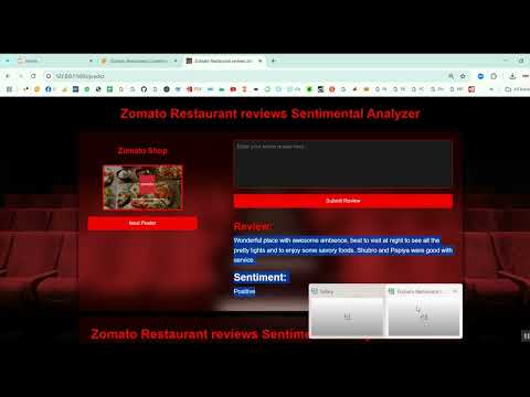
Zomato Restaurant Reviews Sentimental Analyzer in Python Projects
Zomato Restaurant Reviews Sentimental Analyzer in Python Projects
र 1000
⭐ Featured
⭐ Featured
AI & ML Models
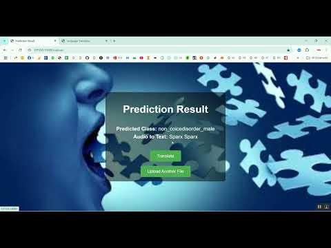
Voice Disorder Prediction using Audio Dataset in Python Projects
Voice Disorder Prediction using Audio Dataset in Python Projects
र 1000
AI & ML Models
Vitamin Deficiency Detection Using Image Processing in Python Projects
Vitamin Deficiency Detection Using Image Processing in Python Projects
र 1000