AI & ML Models
0.0 (0 reviews) • 0 downloads
Brain Tumor Detection 2d 3d Image Classification in Python Projects
र 1000
Brain Tumor Detection 2d 3d Image Classification in Python Projects
Technical Details
Domain : Python
Database : Sqlite
Tools : Anaconda
Run Tools: VS Code
Database : Sqlite
Tools : Anaconda
Run Tools: VS Code
Secure Payment
Instant Download
GST Invoice
24/7 Support
About This Product
Brain Tumor Detection 2d 3d Image Classification in Python Projects
Abstract
Early and accurate detection of brain tumors is essential for effective treatment planning and patient care. This project presents a Brain Tumor Detection System using Python that classifies both 2D MRI slices and 3D volumetric images to identify and categorize brain tumors. The system leverages Convolutional Neural Networks (CNNs) for 2D images and 3D CNN architectures for volumetric MRI scans, extracting spatial and structural features for accurate classification. Python libraries such as TensorFlow/Keras, PyTorch, OpenCV, NumPy, and Matplotlib are used for image preprocessing, model training, evaluation, and visualization. By integrating 2D and 3D image analysis, the system improves diagnostic accuracy, reduces manual workload for radiologists, and supports timely clinical decision-making.
Existing System
In existing systems, brain tumor detection is often performed manually by radiologists using 2D MRI slices, which is time-consuming and subject to human error. Some automated methods focus only on 2D images using conventional image processing techniques such as thresholding, edge detection, or segmentation, but they often fail to capture volumetric and structural tumor information. 3D imaging analysis methods exist but are computationally intensive, require extensive preprocessing, and often use handcrafted features that limit accuracy and generalization. Existing systems struggle to provide an integrated approach that can handle both 2D and 3D data for robust tumor detection.
Proposed System
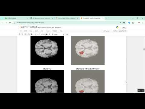
The proposed system introduces a Python-based deep learning framework for 2D and 3D brain tumor detection. MRI scans are preprocessed using noise reduction, normalization, skull stripping, and resizing. For 2D image classification, CNNs extract features from individual slices to detect tumor presence and type. For volumetric MRI data, 3D CNNs analyze the entire scan to capture spatial and structural tumor information. The system uses data augmentation techniques to enhance model generalization. Model performance is evaluated using metrics such as accuracy, precision, recall, F1-score, Dice coefficient, and Intersection over Union (IoU). Visualization tools highlight detected tumor regions on 2D slices and 3D reconstructions. A Jupyter Notebook or Streamlit interface allows clinicians to upload MRI scans, view segmentation results, and receive automated tumor classification. This approach provides a comprehensive, efficient, and scalable solution for brain tumor detection in clinical settings.
Abstract
Early and accurate detection of brain tumors is essential for effective treatment planning and patient care. This project presents a Brain Tumor Detection System using Python that classifies both 2D MRI slices and 3D volumetric images to identify and categorize brain tumors. The system leverages Convolutional Neural Networks (CNNs) for 2D images and 3D CNN architectures for volumetric MRI scans, extracting spatial and structural features for accurate classification. Python libraries such as TensorFlow/Keras, PyTorch, OpenCV, NumPy, and Matplotlib are used for image preprocessing, model training, evaluation, and visualization. By integrating 2D and 3D image analysis, the system improves diagnostic accuracy, reduces manual workload for radiologists, and supports timely clinical decision-making.
Existing System
In existing systems, brain tumor detection is often performed manually by radiologists using 2D MRI slices, which is time-consuming and subject to human error. Some automated methods focus only on 2D images using conventional image processing techniques such as thresholding, edge detection, or segmentation, but they often fail to capture volumetric and structural tumor information. 3D imaging analysis methods exist but are computationally intensive, require extensive preprocessing, and often use handcrafted features that limit accuracy and generalization. Existing systems struggle to provide an integrated approach that can handle both 2D and 3D data for robust tumor detection.
Proposed System
The proposed system introduces a Python-based deep learning framework for 2D and 3D brain tumor detection. MRI scans are preprocessed using noise reduction, normalization, skull stripping, and resizing. For 2D image classification, CNNs extract features from individual slices to detect tumor presence and type. For volumetric MRI data, 3D CNNs analyze the entire scan to capture spatial and structural tumor information. The system uses data augmentation techniques to enhance model generalization. Model performance is evaluated using metrics such as accuracy, precision, recall, F1-score, Dice coefficient, and Intersection over Union (IoU). Visualization tools highlight detected tumor regions on 2D slices and 3D reconstructions. A Jupyter Notebook or Streamlit interface allows clinicians to upload MRI scans, view segmentation results, and receive automated tumor classification. This approach provides a comprehensive, efficient, and scalable solution for brain tumor detection in clinical settings.
Customer Reviews (0)
No reviews yet. Be the first!
Related Products
⭐ Featured

AI & ML Models
Zomato Restaurant Reviews Sentimental Analyzer in Python Projects
Zomato Restaurant Reviews Sentimental Analyzer in Python Projects
र 1000
⭐ Featured
⭐ Featured

AI & ML Models
Voice Disorder Prediction using Audio Dataset in Python Projects
Voice Disorder Prediction using Audio Dataset in Python Projects
र 1000
AI & ML Models
Vitamin Deficiency Detection Using Image Processing in Python Projects
Vitamin Deficiency Detection Using Image Processing in Python Projects
र 1000