AI & ML Models
0.0 (0 reviews) • 0 downloads
Brain Tumor CNN Image Segmentation in Python Projects
र 1000
Brain Tumor CNN Image Segmentation in Python Projects
Technical Details
Domain : Python
Database : Sqlite
Tools : Anaconda
Run Tools: VS Code
Database : Sqlite
Tools : Anaconda
Run Tools: VS Code
Secure Payment
Instant Download
GST Invoice
24/7 Support
About This Product
Brain Tumor CNN Image Segmentation in Python Projects
Abstract
Brain tumor segmentation in medical imaging is a crucial step for diagnosis, treatment planning, and surgical navigation. Manual segmentation of tumors from MRI scans is time-consuming, error-prone, and subject to inter-observer variability. This project, Brain Tumor CNN Image Segmentation in Python, proposes a deep learning approach using Convolutional Neural Networks (CNNs) to automatically segment tumors from brain MRI images. The system uses Python libraries such as TensorFlow/Keras, OpenCV, NumPy, and Matplotlib to preprocess MRI images, train CNN models, and segment tumor regions with high accuracy. By learning hierarchical spatial features, CNNs can distinguish between healthy tissues and tumor regions, producing pixel-wise segmentation masks. The proposed system aims to improve accuracy, reduce diagnosis time, and assist radiologists in medical decision-making.
Existing System
Traditional brain tumor detection and segmentation methods include manual radiologist annotation, thresholding, region-growing, and clustering-based approaches such as K-means or Fuzzy C-means. While these methods can provide initial results, they struggle with complex tumor shapes, low-contrast boundaries, and variability across patients. Machine learning methods with handcrafted features also exist but lack generalization when applied to large datasets. Manual and semi-automated techniques are also time-intensive and prone to subjective bias, limiting their scalability in clinical practice.
Proposed System
The proposed system introduces an automated CNN-based segmentation model for brain MRI scans. The process begins with image preprocessing steps such as grayscale conversion, normalization, skull-stripping, and noise reduction. The preprocessed images are then fed into a CNN architecture (such as U-Net, SegNet, or a custom encoder-decoder CNN) that performs pixel-level classification to generate tumor segmentation masks. The system is trained on publicly available MRI datasets and evaluated using performance metrics like Dice coefficient, Intersection over Union (IoU), accuracy, and sensitivity. Compared to existing systems, this deep learning approach provides higher accuracy, robustness to variations in tumor shape/size, automation, and scalability. The segmented tumor masks can be overlaid on MRI scans to provide radiologists with precise tumor localization and volume estimation.
Abstract
Brain tumor segmentation in medical imaging is a crucial step for diagnosis, treatment planning, and surgical navigation. Manual segmentation of tumors from MRI scans is time-consuming, error-prone, and subject to inter-observer variability. This project, Brain Tumor CNN Image Segmentation in Python, proposes a deep learning approach using Convolutional Neural Networks (CNNs) to automatically segment tumors from brain MRI images. The system uses Python libraries such as TensorFlow/Keras, OpenCV, NumPy, and Matplotlib to preprocess MRI images, train CNN models, and segment tumor regions with high accuracy. By learning hierarchical spatial features, CNNs can distinguish between healthy tissues and tumor regions, producing pixel-wise segmentation masks. The proposed system aims to improve accuracy, reduce diagnosis time, and assist radiologists in medical decision-making.
Existing System
Traditional brain tumor detection and segmentation methods include manual radiologist annotation, thresholding, region-growing, and clustering-based approaches such as K-means or Fuzzy C-means. While these methods can provide initial results, they struggle with complex tumor shapes, low-contrast boundaries, and variability across patients. Machine learning methods with handcrafted features also exist but lack generalization when applied to large datasets. Manual and semi-automated techniques are also time-intensive and prone to subjective bias, limiting their scalability in clinical practice.
Proposed System
The proposed system introduces an automated CNN-based segmentation model for brain MRI scans. The process begins with image preprocessing steps such as grayscale conversion, normalization, skull-stripping, and noise reduction. The preprocessed images are then fed into a CNN architecture (such as U-Net, SegNet, or a custom encoder-decoder CNN) that performs pixel-level classification to generate tumor segmentation masks. The system is trained on publicly available MRI datasets and evaluated using performance metrics like Dice coefficient, Intersection over Union (IoU), accuracy, and sensitivity. Compared to existing systems, this deep learning approach provides higher accuracy, robustness to variations in tumor shape/size, automation, and scalability. The segmented tumor masks can be overlaid on MRI scans to provide radiologists with precise tumor localization and volume estimation.
Customer Reviews (0)
No reviews yet. Be the first!
Related Products
⭐ Featured
AI & ML Models
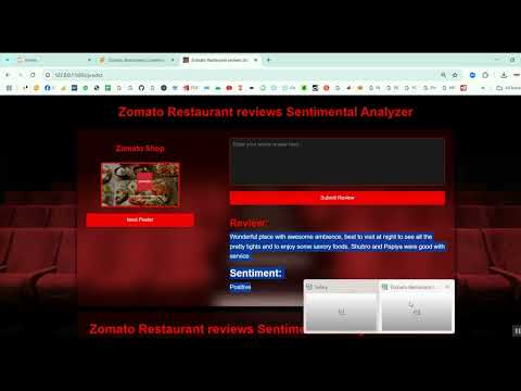
Zomato Restaurant Reviews Sentimental Analyzer in Python Projects
Zomato Restaurant Reviews Sentimental Analyzer in Python Projects
र 1000
⭐ Featured
⭐ Featured
AI & ML Models
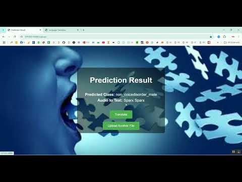
Voice Disorder Prediction using Audio Dataset in Python Projects
Voice Disorder Prediction using Audio Dataset in Python Projects
र 1000
AI & ML Models
Vitamin Deficiency Detection Using Image Processing in Python Projects
Vitamin Deficiency Detection Using Image Processing in Python Projects
र 1000