AI & ML Models
0.0 (0 reviews) • 0 downloads
Brain MRI image Segmentation in Python Projects
र 1000
Brain MRI image Segmentation in Python Projects
Technical Details
Domain : Python
Database : Sqlite
Tools : Anaconda
Run Tools: VS Code
Database : Sqlite
Tools : Anaconda
Run Tools: VS Code
Secure Payment
Instant Download
GST Invoice
24/7 Support
About This Product
Brain MRI image Segmentation in Python Projects
Abstract
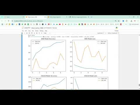
Brain MRI image segmentation is a critical task in medical imaging for diagnosing neurological disorders, tumor detection, and treatment planning. This project presents a Brain MRI Image Segmentation System using Python, which leverages deep learning techniques to automatically segment brain tissues, tumors, or lesions from MRI scans. The system uses Convolutional Neural Networks (CNNs) and U-Net architectures for pixel-level segmentation, providing high accuracy and robust performance. Python libraries including TensorFlow/Keras, OpenCV, NumPy, Pandas, and Matplotlib are used for image preprocessing, model training, evaluation, and visualization. By automating brain MRI segmentation, the system reduces manual workload, improves diagnostic precision, and assists clinicians in planning effective treatments.
Existing System
In existing systems, brain MRI segmentation is primarily performed manually by radiologists, which is time-consuming, prone to human error, and dependent on expertise. Semi-automated approaches using traditional image processing methods such as thresholding, edge detection, region growing, and clustering are limited in handling complex tissue structures, varying intensities, and noisy images. Some machine learning-based methods exist, but they often rely on handcrafted features, which reduces accuracy and generalization across different MRI datasets. Consequently, these methods struggle to provide reliable, fast, and scalable segmentation for clinical and research purposes.
Proposed System
The proposed system introduces a Python-based deep learning framework for brain MRI image segmentation. MRI scans are preprocessed with noise reduction, intensity normalization, skull stripping, and resizing to standard dimensions. A U-Net or similar CNN encoder-decoder architecture is employed to perform pixel-wise segmentation, distinguishing different brain regions, lesions, or tumors. Model performance is evaluated using metrics such as Dice coefficient, Intersection over Union (IoU), accuracy, and precision. Visualization tools in Python, using Matplotlib or Seaborn, highlight segmented regions and overlay results on original images for clinical interpretation. A Jupyter Notebook or Streamlit interface can allow users to upload MRI scans, view segmented outputs, and generate reports for medical evaluation. This approach ensures accurate, automated, and efficient segmentation, supporting clinical decision-making and neuroscience research.
Abstract
Brain MRI image segmentation is a critical task in medical imaging for diagnosing neurological disorders, tumor detection, and treatment planning. This project presents a Brain MRI Image Segmentation System using Python, which leverages deep learning techniques to automatically segment brain tissues, tumors, or lesions from MRI scans. The system uses Convolutional Neural Networks (CNNs) and U-Net architectures for pixel-level segmentation, providing high accuracy and robust performance. Python libraries including TensorFlow/Keras, OpenCV, NumPy, Pandas, and Matplotlib are used for image preprocessing, model training, evaluation, and visualization. By automating brain MRI segmentation, the system reduces manual workload, improves diagnostic precision, and assists clinicians in planning effective treatments.
Existing System
In existing systems, brain MRI segmentation is primarily performed manually by radiologists, which is time-consuming, prone to human error, and dependent on expertise. Semi-automated approaches using traditional image processing methods such as thresholding, edge detection, region growing, and clustering are limited in handling complex tissue structures, varying intensities, and noisy images. Some machine learning-based methods exist, but they often rely on handcrafted features, which reduces accuracy and generalization across different MRI datasets. Consequently, these methods struggle to provide reliable, fast, and scalable segmentation for clinical and research purposes.
Proposed System
The proposed system introduces a Python-based deep learning framework for brain MRI image segmentation. MRI scans are preprocessed with noise reduction, intensity normalization, skull stripping, and resizing to standard dimensions. A U-Net or similar CNN encoder-decoder architecture is employed to perform pixel-wise segmentation, distinguishing different brain regions, lesions, or tumors. Model performance is evaluated using metrics such as Dice coefficient, Intersection over Union (IoU), accuracy, and precision. Visualization tools in Python, using Matplotlib or Seaborn, highlight segmented regions and overlay results on original images for clinical interpretation. A Jupyter Notebook or Streamlit interface can allow users to upload MRI scans, view segmented outputs, and generate reports for medical evaluation. This approach ensures accurate, automated, and efficient segmentation, supporting clinical decision-making and neuroscience research.
Customer Reviews (0)
No reviews yet. Be the first!
Related Products
⭐ Featured

AI & ML Models
Zomato Restaurant Reviews Sentimental Analyzer in Python Projects
Zomato Restaurant Reviews Sentimental Analyzer in Python Projects
र 1000
⭐ Featured
⭐ Featured

AI & ML Models
Voice Disorder Prediction using Audio Dataset in Python Projects
Voice Disorder Prediction using Audio Dataset in Python Projects
र 1000
AI & ML Models
Vitamin Deficiency Detection Using Image Processing in Python Projects
Vitamin Deficiency Detection Using Image Processing in Python Projects
र 1000