AI & ML Models
0.0 (0 reviews) • 0 downloads
Bone X-Ray Jupiter in Python Projects
र 1000
Bone X-Ray Jupiter in Python Projects
Technical Details
Domain : Python
Database : Sqlite
Tools : Anaconda
Run Tools: VS Code
Database : Sqlite
Tools : Anaconda
Run Tools: VS Code
Secure Payment
Instant Download
GST Invoice
24/7 Support
About This Product
Bone X-Ray Jupiter in Python Projects
Abstract
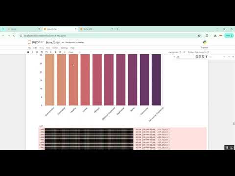
Bone X-ray analysis plays a critical role in diagnosing fractures, osteoporosis, and other skeletal disorders. This project presents a Bone X-Ray Analysis System using Python in Jupyter Notebook, which leverages image processing and deep learning techniques to automatically detect and classify bone abnormalities from X-ray images. The system uses Convolutional Neural Networks (CNNs) to extract features and predict conditions such as fractures, bone density variations, or deformities. Python libraries including OpenCV, TensorFlow/Keras, NumPy, Pandas, and Matplotlib are used for preprocessing, model training, evaluation, and visualization. By automating X-ray analysis, the system reduces manual workload, improves diagnostic accuracy, and provides a scalable tool for clinical applications.
Existing System
In existing systems, bone X-ray analysis is primarily performed manually by radiologists, who visually inspect images for fractures, bone density abnormalities, or structural defects. This process is time-consuming, subjective, and prone to inter-observer variability. Some semi-automated tools exist to assist in highlighting regions of interest or measuring bone density, but they often require manual intervention and expertise. Traditional image processing and machine learning approaches using handcrafted features struggle to generalize across different image qualities, patients, and X-ray machines, limiting accuracy and scalability.
Proposed System
The proposed system introduces a Python-based deep learning framework for automated bone X-ray analysis within a Jupyter Notebook environment. Bone X-ray images are preprocessed using techniques such as grayscale conversion, noise reduction, histogram equalization, and segmentation to enhance key structures. Features are extracted automatically using a CNN architecture such as ResNet, VGG, or DenseNet, and used to classify X-rays into categories like normal, fractured, or osteoporotic. Model performance is evaluated using metrics such as accuracy, precision, recall, F1-score, and confusion matrix. Interactive visualization within Jupyter Notebook allows clinicians to view X-ray images, predicted classifications, and highlighted abnormal regions. This approach ensures accurate, efficient, and reproducible bone X-ray analysis suitable for hospitals, research centers, and educational purposes.
Abstract
Bone X-ray analysis plays a critical role in diagnosing fractures, osteoporosis, and other skeletal disorders. This project presents a Bone X-Ray Analysis System using Python in Jupyter Notebook, which leverages image processing and deep learning techniques to automatically detect and classify bone abnormalities from X-ray images. The system uses Convolutional Neural Networks (CNNs) to extract features and predict conditions such as fractures, bone density variations, or deformities. Python libraries including OpenCV, TensorFlow/Keras, NumPy, Pandas, and Matplotlib are used for preprocessing, model training, evaluation, and visualization. By automating X-ray analysis, the system reduces manual workload, improves diagnostic accuracy, and provides a scalable tool for clinical applications.
Existing System
In existing systems, bone X-ray analysis is primarily performed manually by radiologists, who visually inspect images for fractures, bone density abnormalities, or structural defects. This process is time-consuming, subjective, and prone to inter-observer variability. Some semi-automated tools exist to assist in highlighting regions of interest or measuring bone density, but they often require manual intervention and expertise. Traditional image processing and machine learning approaches using handcrafted features struggle to generalize across different image qualities, patients, and X-ray machines, limiting accuracy and scalability.
Proposed System
The proposed system introduces a Python-based deep learning framework for automated bone X-ray analysis within a Jupyter Notebook environment. Bone X-ray images are preprocessed using techniques such as grayscale conversion, noise reduction, histogram equalization, and segmentation to enhance key structures. Features are extracted automatically using a CNN architecture such as ResNet, VGG, or DenseNet, and used to classify X-rays into categories like normal, fractured, or osteoporotic. Model performance is evaluated using metrics such as accuracy, precision, recall, F1-score, and confusion matrix. Interactive visualization within Jupyter Notebook allows clinicians to view X-ray images, predicted classifications, and highlighted abnormal regions. This approach ensures accurate, efficient, and reproducible bone X-ray analysis suitable for hospitals, research centers, and educational purposes.
Customer Reviews (0)
No reviews yet. Be the first!
Related Products
⭐ Featured

AI & ML Models
Zomato Restaurant Reviews Sentimental Analyzer in Python Projects
Zomato Restaurant Reviews Sentimental Analyzer in Python Projects
र 1000
⭐ Featured
⭐ Featured
AI & ML Models
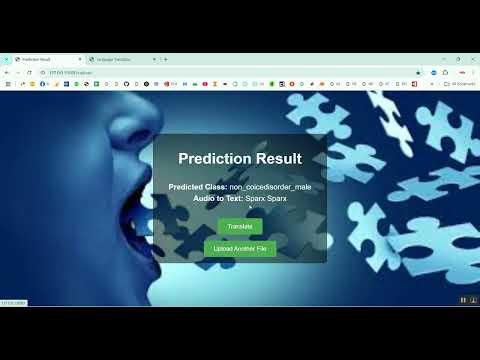
Voice Disorder Prediction using Audio Dataset in Python Projects
Voice Disorder Prediction using Audio Dataset in Python Projects
र 1000
AI & ML Models
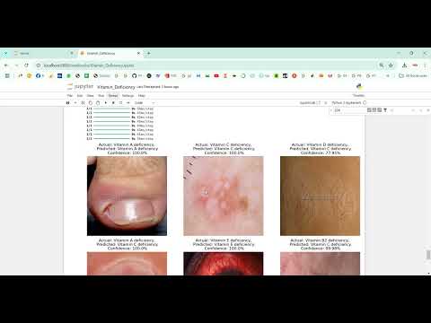
Vitamin Deficiency Detection Using Image Processing in Python Projects
Vitamin Deficiency Detection Using Image Processing in Python Projects
र 1000